URSL Method

சிறுநீரகக் கல் நோய்

நம்முடைய சிறுநீரகங்கள் வயிற்றின் மேல் பகுதியில் இரண்டு புறமும் ஒவ்வொன்றாக இருக்கின்றன. சிறுநீரகத்தில் உற்பத்தியாகும் சிறுநீர் 'யுரீடர்' என்ற குழாய்கள் மூலம் சிறுநீர்ப்பைக்கு வந்து சேர்கின்றது. சிறுநீர்ப்பையில் சிறுநீர் சேகரிக்கப்படுகிறது. அங்கிருந்து மூத்திரக் குழாய் மூலம் சிறுநீர் வெளியேற்றப்படுகிறது.

சிறுநீரக மண்டலத்தில் கற்கள் உண்டாவதால் ஏற்படும் அறிகுறிகள் என்ன?

சிறுநீரக மண்டலத்தில் கற்கள் உண்டாவதால் ஏற்படும் அறிகுறிகள் கல் எந்த இடத்தில் இருக்கிறது என்பதைப் பொறுத்தது.

சிறுநீரகக் கற்கள்

கற்கள் சிறுநீரகத்துக்கு உள்ளே இருக்கும்போது லேசான வலியே இருக்கும். சில சமயங்களில் காய்ச்சல் ஏற்படலாம். சிறுநீரில் ரத்தம் கலந்து வரலாம்.

யுரீட்டர் கற்கள்

கற்கள் யுரீட்டரில் செல்லும்போது மிகக் கடுமையான வலி ஏற்படும். விட்டு, விட்டு பிசைவது போல இருக்கும். யுரீட்டரின் மேற்பகுதியில் இருக்கும்போது முதுகிலிருந்து வயிற்றின் முன்பக்கம் பரவிவரும். நடுவில் இருக்கும் போது வயிற்றின் பக்கவாட்டில் இருந்து அடிவயிற்றுக்குப் பரவும்.  இன்னும் கீழே வந்தால் சிறுநீர் கழிக்கும் போது எரிச்சல் ஏற்படும். சிறுநீர் வரும் வழி எங்கும் வலிக்கும்.

மூத்திரப் பையில் கற்கள்

மூத்திரப் பையில் சிறிய கற்கள் உண்டாகும் போது அடிவயிற்றில் லேசான வலி இருக்கும். சிறுநீர் கழிக்கும் போது எரிச்சல் ஏற்படும். சிறுநீர் வருவது அடைத்துக்கொண்டு வலி ஏற்படலாம். சில சமயம் சிறுநீர் கழிக்க முடியாமல் போகலாம்.

மூத்திரக் குழாயில் கற்கள்

கல் மூத்திர குழாயில் இருக்கும்போது சிறு நீர் பாய்ச்சுவது கடினமாக இருக்கும். சுத்தமாக சிறுநீர் கழிக்க முடியாமல் போகலாம்.

சிறுநீரக கற்களுக்கான மருத்துவங்கள் என்ன?

பொதுவாக சிறிய கற்கள் இயற்கையாகவே சிறுநீர் வழியாக வெளியே வந்துவிடும். அப்படிப்பட்ட க் கற்களுக்கு ஆன்ட்டிபயாட்டிக் மருந்துகளும், வலி மருந்துகளும் கொடுத்தால் போதுமானது. பொதுவாக 7 மில்லி மீட்டருக்குக் குறைவான கற்கள் எளிதாக வந்துவிடும். 

7 மில்லி மீட்டரை விட பெரிய கற்கள் கவனமாக பார்க்கப்பட வேண்டியவை. சில சமயம் 12 மில்லி மீட்டர் அளவுள்ள கற்கள் கூட சிறுநீரிலேயே வெளியே வந்துவிடலாம். எனவே எந்தக் கல்லாக இருந்தாலும் மருந்து மூலம் கல் வருவதற்கு வழி இருக்கிறதா என்று பார்த்து, முடியவில்லை என்கிற பொழுது அறுவை சிகிச்சைப் பற்றி நினைக்க வேண்டும்.

சிறுநீரக கற்களை எடுப்பதற்கான சிகிச்சைமுறைகள்

இ.எஸ்.டபிள்யூ.எல்.

இந்த முறையில் சிறுநீரகத்திற்கு உள்ளே இருக்கும் கற்களை அறுவை சிகிச்சை இல்லாமல் மயக்க மருந்து இல்லாமல் உடம்புக்கு வெளியே இருந்து தரப்படும் கதிர்கள் மூலம் உடைக்கலாம். உடைந்த கற்கள் சிறுநீர் வெளியே வந்துவிடும். பொதுவாக 10 மில்லி மீட்டர் வரையிலான சிறுநீரகக் கற்களை இப்படி உடைக்கலாம்.

யுரிடரோஸ்கோபி

சிறுநீரக உள்நோக்கி மூலம் கற்களை உடைப்பது

இந்த முறையில் யுரீடர் குழாயில் உள்ள கற்களை உடைக்கலாம். முதுகில் ஊசி செலுத்தி அல்லது முழுமையான மயக்கம் கொடுத்து சிறுநீரக மண்டலத்திற்கு உள்ளாக சுண்டு விரலில் நான்கில் ஒரு அளவு இருக்கும் உள் நோக்கிக் கருவியை சிறுநீர் பாதை வழியாகச் செலுத்தி கல்லை பார்த்து கல்லை உடைப்பது. கற்களை உடைப்பதற்கு லேசர் அல்லது சுத்தியல் போன்ற கருவி பயன்படுத்தப்படும். கல்லை உடைத்த பிறகு ஸ்டென்ட் ஏற்படும் குழாய் உள்ளே வைக்கப்படும். குழாயைச் சுற்றி உடைந்த கல் துகள்கள் வெளியில் வந்து விடும். ஸ்டென்ட் குழாய் மூன்று வாரம் உள்ளே இருக்கும்.

நவீன வளையக்கூடிய கருவிகள் மூலம் சிறுநீரகத்திற்கு உள்ளே எந்த மூலையில் இருக்கிற கற்களையும் கூட லேசர் மூலம் உடைத்து விட முடியும்.

சிஸ்டோலிதோடிரிப்ஸி

இந்த முறையின் மூலம் சிறுநீரகத்தில் உள்ள கற்களை உடைத்து எடுத்துவிட முடியும். உள்நோக்கி கருவியின் மூலம் மூத்திரப்பை உள்ளே கல்லை நேராகப் பார்த்தேன் அதனை பொடி செய்து,  வெளியே எடுத்துவிட முடியும்.

பெர்குடேனியஸ் நெஃப்ரோலிதோடிரிப்ஸி

இந்த முறையில் எக்ஸ்-ரே கருவிகள் துணையோடு நம்முடைய கிட்னியை நேராக துளையிட்டு அந்த துளையின் மூலம் கிட்னியின் உள்ளே, உள் நோக்கியைச் செலுத்த வேண்டும். உரிய கருவிகள் மூலம் கல்லை உடைத்து எல்லாத் துகள்களையும் வெளியே எடுக்க வேண்டும்.

இந்த அறுவை சிகிச்சைகளின் பக்க விளைவுகள் என்ன?

பொதுவாக உள்நோக்கிக் கருவிகளின் மூலம் செய்யப்படும் அறுவைச் சிகிச்சைகள் பாதுகாப்பானவை. இருப்பினும் கிருமிகள் தொற்று, அதனால் ஏற்படும் பிரச்சினைகள், மயக்க மருந்து கொடுப்பதால் ஏற்படும் பிரச்சினைகள் போன்ற பிரச்சினைகள் ஏற்படலாம். சிறுநீரகத்தை நேராக துளையிடும் போது அதிக ரத்தப்போக்கு ஏற்பட்டு ரத்தம் வீணாகலாம். அப்படிப்பட்டவர்களுக்கு ரத்தம் செலுத்த வேண்டிவரும். சில சமயங்களில் உள்நோக்கிக் கருவிகள் சென்றடைய முடியாத இடங்களில் கல் இருந்தால் அறுவைச் சிகிச்சை தோல்வி அடைந்துவிடும்.

வழக்கமான, திறந்த அறுவை சிகிச்சை

சில சமயங்களில் மிகப் பெரிய கற்களை வயிற்றைக் கிழித்து எடுக்க வேண்டும்.

சிறுநீரகக் கற்கள் வராமல் தடுப்பது எப்படி?

சிறுநீரகக் கற்கள் வராமல் தடுப்பதற்கு, கற்கள் ஏன் உண்டாகின்றன என்பதை அறிந்து கொள்ள வேண்டும். நம்முடைய சிறுநீரகங்களின் பணி, உடலுக்குத் தேவையில்லாத உப்புக்களைக் கரைப்பது. அப்படிக் கரைக்கும் போது சில சிறுநீரகங்கள் முழுமையாகக் கரைக்காமல், கண்ணுக்குத் தெரியாது திப்பிகளை விட்டுவிடுகின்றன. இந்தத் திப்பிகள் இணைந்து கற்களாக மாறிவிடுகின்றன. கரைக்கும் திறன் என்பது ஒவ்வொருவருக்கும் மாறுபடும். சில பேருடைய சிறுநீரகங்கள் இயற்கையாகவே கரைக்கும் திறன் குறைவாகப் பெற்றிருக்கும். இதை வியாதி என்று கூறுவதைவிட அவருடைய உடல்வாகு என்று சொல்வதே சரி. சில குறிப்பிட்ட உடல் உபாதைகள் இன் போதும் சிறுநீரகக் கற்கள் தோன்றும். உதாரணமாக பேரா தைராய்டு நாளமில்லா சுரப்பிகள் அதிகமாக வேலை செய்வதாலும் உடலில் யூரிக் ஆசிட் அதிகமாக இருப்பதாலும் கற்கள் உண்டாகலாம்.

கற்கள் உண்டாவது குறிப்பிட்ட ஒரு உப்பினை அதிகமாக உண்பதால் அல்ல. எனவே நாங்கள் எந்த விதமான உணவுப் பத்தியமோ அல்லது உணவு முறைமோ பரிந்துரைப்பது இல்லை. 

இருப்பினும் தாராளமாக நீர் குடிப்பதால் கற்கள் சிறியதாக இருக்கும்போதே வெளியே வரும் வாய்ப்பு இருக்கின்றது.

Kidney Stones

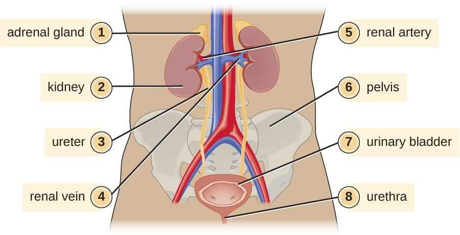
This is our urinary system. Kidneys are in the upper part of our abdomen. The urine secreted by the Kidneys are carried by tubes called ureters to the urinary bladder, where urine is stored. From the bladder urine is passed through urethra.

What are the symptoms of stone disease?

The symptoms of stone disease depend of the location of the stone.

Kidney stones

When stones are in the Kidney they cause a dull, continuous pain. They may produce fever sometimes. Rarely patient may pass blood in urine.

Ureteric Stones

When the stones pass through the ureter they cause severe pain called ureteric colic. This is a severe colicky pain that may radiate from loin to groin. The pain may go to scrotum and penis also. When the stone is in the lower part of the ureter patient will have burning sensation in the urethra and while passing urine.

Bladder stones

Bladder stones produce dull lower abdominal pain. Patient will also have burning sensation while passing urine. Sometimes stones block the passage of urine. Patient may difficulty to pass urine or may be totally not able to pass urine.

Urethral stone

Stones in the urethra can stop urine flow and patient may present with inability to pass urine.

What are the treatments for stone disease?

Small stone are usually passed in urine. They are treated with painkillers, antibiotics and other supportive drugs. It is usually considered that stone of less than 7 millimeters are passed easily

Stones bigger than 7 mms need to be removed if they are not passed in urine. They will need surgical removal

What are the methods for removal of stone:

  • ESWL (Extracorporeal Shockwave Lithotripsy)
  • Cystolithotripsy
  • Ureterorenoscopy
  • Percutaneous Nephrolithotomy
  • Open Surgery

ESWL (Extracorporeal Shockwave Lithotripsy)

ESWL (Extracorporeal Shockwave Lithotripsy) is a method where shockwaves are produced by a machine outside our body and they are beamed over the stone using x ray localization. These shockwaves powder the stone and the powder is passed in urine. There is no anesthesia

Cystolithotripsy

Here an endoscopic instrument called Cystoscope is passed into the bladder and the stone is broken into pieces

Ureterorenoscopy

Here the endoscopic instrument called Ureterorenoscope is passed through the urethra and into the Ureter and the stone is broken using mechanical or laser stone breaking instruments. After breaking the stone, a thin tube called Double J stent is left in the ureter to facilitate passage of broken stones.

Modern flexible ureteroscopes can reach all corners of the Kidney and stones anywhere can be powdered.

Percutaneous Nephrolithotomy

In this method, the Kidney is directly punctured from outside and the passage is widened. A plastic tube is passed to the Kidney itself. Through this tube an endoscopic instrument called Nephroscope is passed and the stone is broken and bits removed. After removal of stone a thin tube called Double J stent is left in the ureter to facilitate passage of any tiny left-over bits of stone.

Open Surgery

The standard open surgery using long incisions to cut the tissues and directly remove the stone from Kidney, ureter or bladder is occasionally necessary for huge stones

What are the complications of stone surgeries?

The endoscopic Urological surgeries are generally safe. However, complications related to infection and anesthesia may happen. Sometimes surgery can fail because the stone is inaccessible to endoscopic instruments. PCNL, where the Kidney is directly punctured can cause excessive bleeding and transfusion may be needed. Rarely, procedures to arrest bleeding may be necessary.

How to prevent further stone formation?

To know how to prevent stone formation, we should understand why stones are formed. The function of our Kidneys is to dissolve unwanted salts from our body and pass them as urine. Urine is nothing but a solution of unwanted salts. While doing this job some Kidneys do not dissolve stones totally. They leave crystals which can only be seen by microscope. These crystals aggregate to form stones. The capacity to dissolve is constitutional. Some conditions like excessive secretion of Paratharmone, a hormone and Hyperuricemia can also cause stone formation.

Stone formation is not due to excessive consumption of certain salts or minerals. Hence we do not advice any food restriction to prevent stone formation. It is advised to consume sufficient water so that stone are passed in urine when they are small in size.